The Nervous System
Function of the Nervous System
The nervous system is the major controlling, regulatory, and communicating system in the body. All mental activity including thought, learning, and memory occurs here.
The nervous system too, is composed of organs, principally the brain, spinal cord, nerves, and ganglia.
The function of the nervous system is critical for every day living and functioning, therefore it is imperative you are familiar with the possible signs and symptoms that could signify injury or dysfunction of any part of this system.
The principle functions of the nervous system are:
- Receive stimuli from outside and inside the body. Analysed and the appropriate coordinated response produced
- Convey impulses from the brain that may stimulate or depress activity in muscular, glandular and other tissues.
- Integrate the many functions carried out by individual organs, tissues and cells.
System Organisation
The nervous system as a whole is divided into two subdivisions:
- The Central Nervous System (CNS). The CNS consists of the brain and spinal cord.
- Peripheral Nervous System (PNS).Twelve pairs of cranial nerves. Thirty-one pairs of spinal nerves. The autonomic nervous system.
Components of the Nervous System
Nervous Tissue
There are two main types of cells in nerve tissue. The actual nerve cell is the neuron. It is the "conducting" cell that transmits impulses and the structural unit of the nervous system. The other type of cell is neuroglia, or glial, cell. The word "neuroglia" means "nerve glue." These cells are nonconductive and provide a support system for the neurons. They are a special type of "connective tissue" for the nervous system.
The Brain
the brain lies within the cranial cavity. It is continuous with the spinal cord and is divided into three main parts:
- Cerebrum
- Cerebellum
- Brain Stem
Cerebrum
The cerebrum is the part of the brain that receives and processes conscious sensation, generates thought, and controls conscious activity. It is the uppermost and largest part of the brain, and is divided into left and right hemispheres.
Each cerebral hemisphere is divided into five lobes, four of which have the same name as the bone over them: the frontal lobe, the parietal lobe, the occipital lobe, and the temporal lobe. A fifth lobe, the insula or Island of Reil, lies deep within the lateral sulcus.
Cerebellum
The Cerebellum is a cauliflower-shaped section of the brain located in the hindbrain, at the bottom rear of the head, directly behind the pons. The cerebellum is a complex system mostly dedicated to the intricate coordination of voluntary movement, including walking and balance. Damage to the cerebellum leaves the sufferer with a gait that appears drunken and is difficult to control.
In Short, responsible for:
- Balance
- Muscle Coordination
- Muscle Tone
Brain Stem
The brain stem is the part of the brain continuous with the spinal cord and comprising the medulla oblongata and pons and midbrain.
-
Medulla Oblongata -
- Controls Autonomic Functions
- Relays Nerve Signals Between the Brain and Spinal Cord
-
Pons Varolii -
- Arousal
- Assists in Controlling Autonomic Functions
- Relays Sensory Information Between the Cerebrum and Cerebellum
- Sleep
-
Midbrain (Mesencephalon) -
- Controls Responses to Sight
- Eye Movement
- Pupil Dilation
- Body Movement
- Hearing
Meninges - The meninges cover the brain and spinal cord. The meninges consist of the pia mater, dura mater and the arachnoid.
Function: Protects Cranial Nerves and Spinal Cord
Dura Mater - The outer layer, the dura mater, is tough white fibrous connective tissue.
Arachnoid Mater - The middle layer of meninges is arachnoid, a thin layer resembling a cobweb with numerous threadlike strands attaching it to the innermost layer. The space under the arachnoid, the subarachnoid space, is filled with cerebrospinal fluid (CSF) and contains blood vessels.
Pia Mater - The pia mater is the innermost layer of meninges. This thin, delicate membrane is tightly bound to the surface of the brain and spinal cord and cannot be dissected away without damaging the surface.
Cerebrospinal Fluid
Function - clear, colourless liquid that fills the ventricles (cavities) of the brain and the spinal cord, surrounds them as well, and acts as a lubricant and a mechanical barrier against shock.
Cranial Nerves
The cranial nerves are 12 pairs of nerves that can be seen on the bottom surface of the brain. Some of these nerves bring information from the sense organs to the brain; other cranial nerves control muscles; other cranial nerves are connected to glands or internal organs such as the heart and lungs. These nerves are listed on the left with their name and corresponding number.
Spinal Cord
The spinal cord extends from the foramen magnum at the base of the skull to the level of the first lumbar vertebra. The cord is continuous with the medulla oblongata at the foramen magnum. Like the brain, the spinal cord is surrounded by bone, meninges, and cerebrospinal fluid.
Two Main Functions:
- Serving as a conduction pathway for impulses going to and from the brain. Sensory impulses travel to the brain on ascending tracts in the cord. Motor impulses travel on descending tracts.
- Serving as a reflex centre. The reflex arc is the functional unit of the nervous system. Reflexes are responses to stimuli that do not require conscious thought and consequently, they occur more quickly than reactions that require thought processes. For example, with the withdrawal reflex, the reflex action withdraws the affected part before you are aware of the pain. Many reflexes are mediated in the spinal cord without going to the higher brain centres.
Peripheral Nerves
Two types of Peripheral Nerve:
- Sensory
- Motor
The peripheral nervous system consists of the nerves that branch out from the brain and spinal cord. These nerves form the communication network between the CNS and the body parts. The peripheral nervous system is further subdivided into the somatic nervous system and the autonomic nervous system.
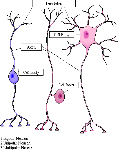
Structure of a Neuron
A neuron contains bundles of nerve fibres, either axons or dendrites, surrounded by connective tissue.
- Sensory or Afferent nerves contain only afferent fibres, long dendrites of sensory neurons. These convey information from tissues and organs to the central nervous system
- Motor nerves have only efferent fibres, long axons of motor neurons. Transmit signals from the the central nervous system
- Mixed nerves contain both types of fibres.
Neurons are made up of three basic parts:
- Cell body or Soma - The central part of the cell between the dendrites and the axon. The nucleus is located here.
- Axon - This structure carries nerve signals away from the neuron. Axons are covered by a protective covering called the myelin sheath. This sheath acts as an insulation thus improving conduction of nerve impulses down the axon.
- Dendrites - Conduct nerve impulses towards the neurons cell body. Unlike axons above, dendrites are not covered by any outer covering

Classification of Neurons:
- Bipolar - A single axon and dendrite arise at opposite poles of the cell body. Found only in sensory neurons, such as in the Retina, Olfactory and auditory systems
- Unipolar - A single process or fibre which divides close to the cell body into two main branches (axon and dendrite). These are also sensory neurons
- Multipolar - Has numerous cell processes (an axon and many dendrites). These are Motor neurons
Nerve cells are connected to each other at a junction known as a synapse, where the end branches of an axon and the dendrites of another neuron are in close proximity to each other but never make direct contact. Information is transmitted chemically between the two neurons at the synaptic cleft by way of diffusion.
Function of the Autonomic Nervous System
The autonomic nervous system is a visceral efferent system, which means it sends motor impulses to the visceral organs. It functions automatically without conscious effort, to innervate smooth muscle, cardiac muscle, and glands. It is concerned with heart rate, breathing rate, blood pressure, body temperature, and other visceral activities that work together to maintain homeostasis.
The autonomic nervous system has two parts, the sympathetic division and the parasympathetic division. Many visceral organs are supplied with fibres from both divisions. In this case, one stimulates and the other inhibits. This relationship serves to help maintain homeostasis.
- Sympathetic Nerves - Stimulating and quickening effect on the heart, circulatory and respiratory systems, but inhibits peristalsis. This system activates what is termed the fight or take flight response
- Parasympathetic Nerves - These have the opposite effect by slowing the heart, reduces circulation and respiration but stimulates digestion. "Rest and Digest".
A Summary of the Effects by the Sympathetic and Parasympathetic Nerves
| Structure | Sympathetic | Parasympathetic |
| Heart | Accelerates Heartbeat | Slows Heartbeat |
| Eyes | Pupil Dilation | Pupil Constriction |
| Lungs | Relaxes Airway | Constricts Airways |
| Stomach | Inhibits Digestion | Stimulates Digestion |